Reference Peak Normalization
What it is
Reference Peak Normalization rescales ion intensities across all pixels relative to a selected reference peak. This corrects for pixel-to-pixel signal variation caused by factors unrelated to biology, such as matrix heterogeneity, tissue thickness variation, or ion suppression, producing a more accurate and spatially comparable ion image.
When to use it
- When your ion images show spatial intensity patterns you suspect reflect sample preparation artifacts rather than genuine biology.
- When you have a known internal standard or a stable endogenous peak that should be uniformly distributed across the tissue and can serve as a reliable normalization reference.
How to use it
Here's a short video showing how to apply reference peak normalization:
What results look like
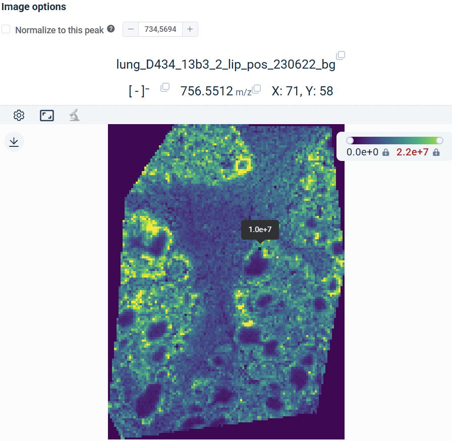
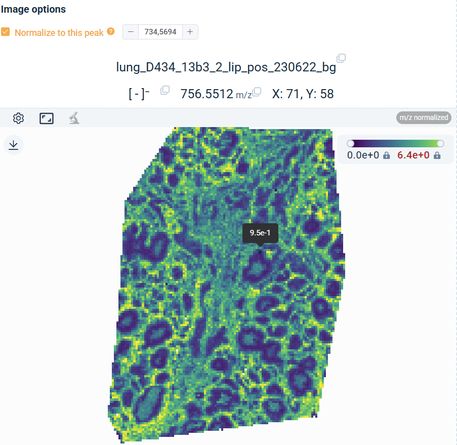
Once a reference peak is selected, the Image Viewer re-renders each ion image as the intensity ratio of the displayed ion relative to that of the reference peak. The reference m/z value is shown alongside the Normalize to this peak toggle. As you browse different m/z values or formulas, all ion images continue to render as ratios to the same reference peak.