Optical Image Overlay
What it is
The optical image overlay tool lets you upload an optical image and align it to your ion images using linear transformations — rotation, scaling, and translation. Once aligned and saved, the optical image is available as a background layer in the annotation page viewer.
When to use it
This tool is useful when you have a complementary image of the same tissue section, such as an H&E stain, a light microscopy image, or a slide layout. Overlaying the optical image on your ion images provides spatial context for interpreting ion localization in biologically meaningful regions, such as tissue layers, tumor margins, or annotated anatomical structures.
How to use it
Instructions for aligning your ion and optical images are available directly on the tool page. Here's a step-by-step video showing how to access and use the optical image overlay tool:
What results look like
Once you are satisfied with the alignment, submit it to save it to your dataset. Returning to the annotation page, the ion image viewer will now display both images simultaneously, with an opacity slider to control the relative transparency of each layer.
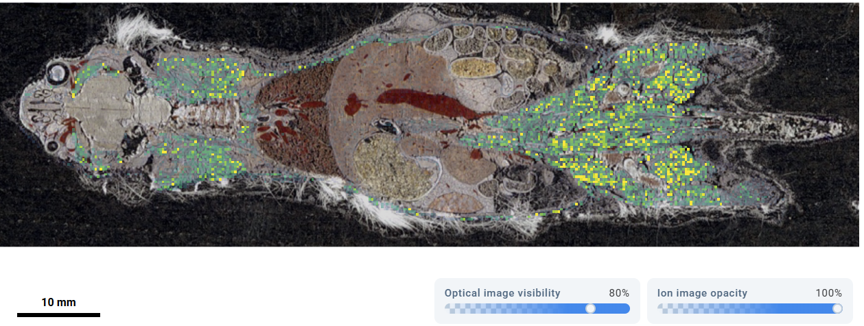 Ion image overlaid on an optical image, with the opacity slider controlling the blend between layers. Source: dataset