ROI Selection
What it is
ROI Selection lets you draw and label regions of interest directly on the ion image viewer. Each ROI defines a spatial boundary around a tissue area you want to isolate for exploration, comparison, or use as input for downstream differential analysis.
When to use it
- When you have prior knowledge of tissue structure and want to define regions based on histology or an overlaid optical image.
- When you want to isolate a specific area, such as a tumor core, a tissue layer, or an anatomical structure — for focused exploration.
- When you want to compare regions you have defined yourself, rather than relying on data-driven segmentation.
How to use it
Here's a short video showing how to manually select and define ROIs:
What results look like
Drawn ROIs appear as labeled boundary overlays on the ion image viewer, each outlined in a distinct color. All defined ROIs are listed in the ROI panel alongside the viewer, where each region can be renamed, recolored, or removed.
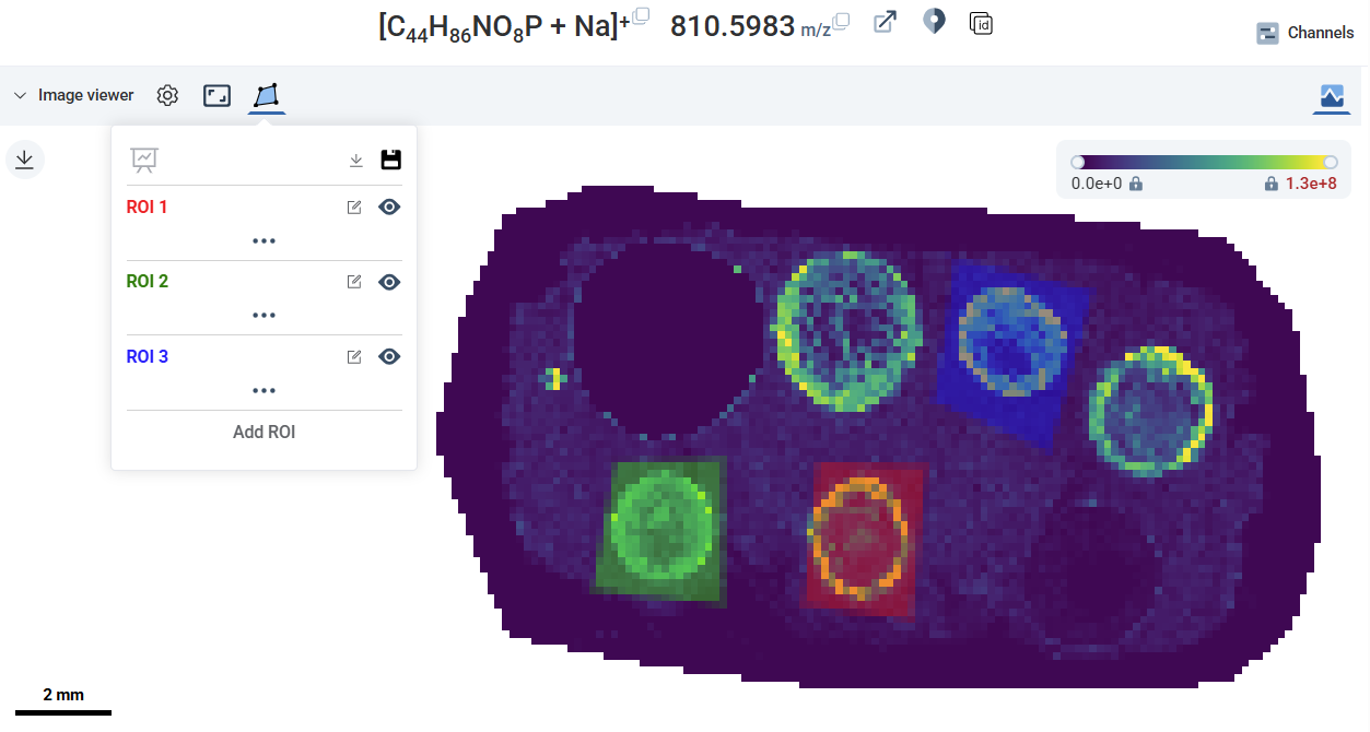 Ion image viewer with multiple ROI boundaries drawn and color-coded, and the ROI panel listing each region with its label and edit controls. Source: dataset